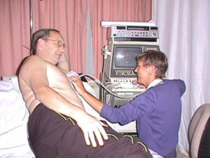
One of my ultra sound scans taking place (me looking like a beached whale)
Does it hurt? No! not a bit.
This is a quick and easy way to look at the valves and chambers of the heart, as well as the blood flow.
The machine they use is much the same as the one they would use to scan pregnant women (not that I am pregnant). First they apply a small amount of gel to your chest, then with the probe up against your chest, they look at your heart. Then using the screen measure your heart, valves and generally check it over. 10/15 minutes and it’s all over, I actually like having it done as you can also see the screen and your own heart beating.
Leslie has done at least 20 or more on me over the passed 5 years and I now count her as a friend, she was my first visitor after the operation, she came down to I.C.U. to see me.
The Official Word
ABOUT YOUR ECHOCARDIOGRAM
An echocardiogram ( or “echo”) is a totally safe and painless ultrasound examination.
It uses high frequency sound waves to form a picture of the valves and chambers of your heart. No special preparation is required for this examination.
Shortly after your arrival in the echo department, you will be shown into a curtained cubicle and asked to remove your clothing above the waist. Should you require assistance, please ask the technician. You will then be asked to lie on a couch on your left side, with your left arm up on the pillow, under your head. If this position is difficult for you, don’t worry it is important that you are as comfortable as possible, and we will try our best to ensure that you are, as you may be there for a little while. An examination can take up to an hour, although the average is between 10 and 20 minutes. During this time, it helps the technician if you can keep as still and quiet as possible.
Once you are settled, we may put sticky electrode pads on your arms or chest so that we can do a basic ECG. A small quantity of gel will then be put on your chest (which might feel a bit cold), and a probe will slide over your skin, please tell the doctor or technician if they press too hard. The probe, or transducer, allows us to see images of your heart on the screen. These images are recorded, so that they can be reviewed later. We may also do a Doppler examination, to give us more information on the flow of blood through the valves of your heart. You will hear this as a “swishing” sound, rather like an old washing machine.
Sometimes, we have doctors or technicians in the department learning this specialized technique. It is important for them to watch or carry out examinations, depending on their stage of training. They will ask your permission before doing this, do not be afraid to refuse if you wish to, similarly if you prefer a male/female technician you should make this known.
When the examination is finished, you will be given some tissue to wipe the gel from your chest; when you are ready you may dress and leave. We will not be able to give you the results of your echo immediately, as the traces have to be analysed. However, as soon as the report is available, it will be sent to your doctor and he/she will discuss the results with you.
If you have any comments or suggestions on how we can improve our echo service, please contact one of the staff.